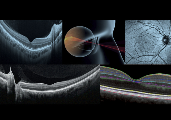
Fast and Easy Acquisition with Incredible Detail
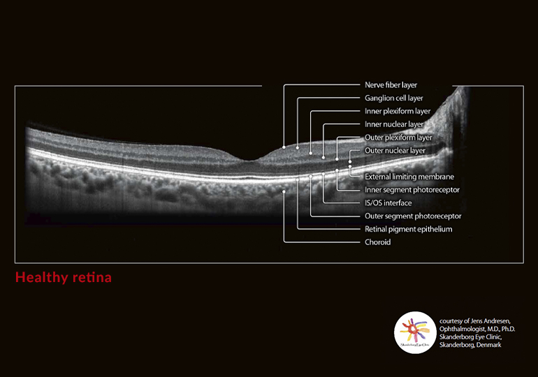
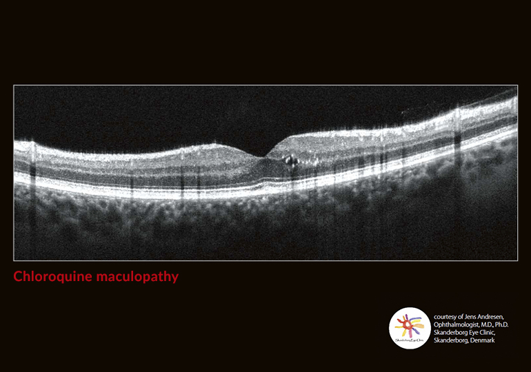
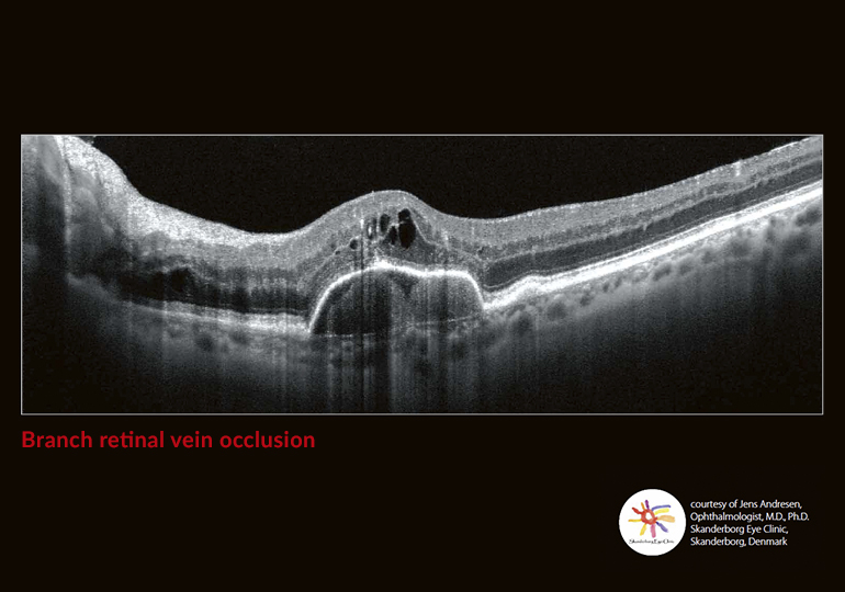
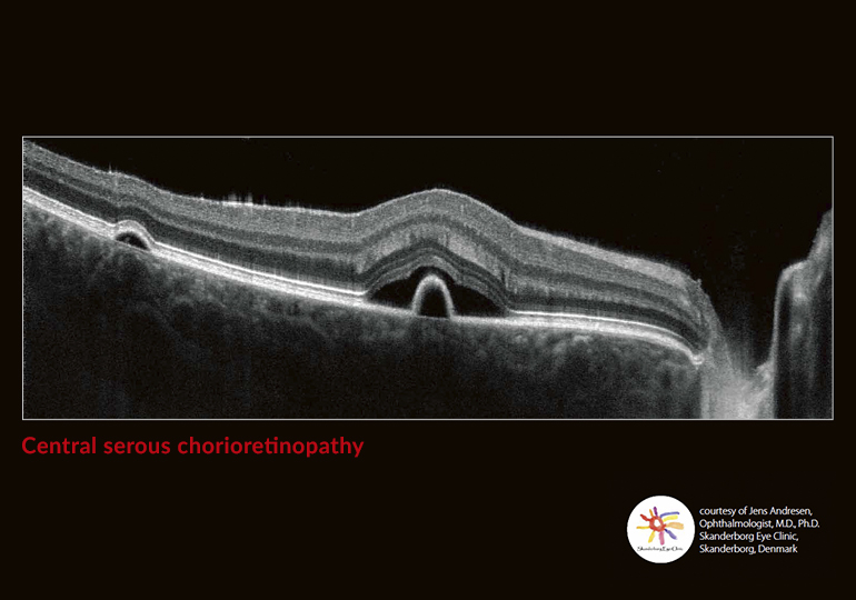
With a digital resolution of up to 1.6μm, the system enables excellent differentiation of structures and individual layers of the retina. High scanning speed of 70,000 A-scans/s enables very short examination times of usually about two seconds, resulting in less motion artefacts and increased patient comfort.